Short answer
Diabetic retinopathy is a disease that develops when diabetes affects the retina and its blood vessels. It may cause no symptoms at first; as it progresses, vessel leakage, bleeding, blockage and new fragile vessel formation can develop. Untreated, it can lead to serious vision loss. Early detection is therefore important for timely action on changes that threaten vision.
Diabetic retinopathy is a disease that occurs when diabetes affects the retina and the retinal blood vessels at the back of the eye. It can progress without causing any symptoms at first. For this reason, normal vision does not necessarily mean that the retina is healthy.
As the disease progresses, leakage, bleeding and blockages can develop in the retinal blood vessels. In more advanced stages, new and fragile blood vessels may form in areas of the retina that are deprived of oxygen. If left untreated, these changes can lead to complications that may cause serious vision loss. Current guidelines emphasize that detecting diabetic retinopathy early is important so that vision-threatening changes can be addressed in time.
How does diabetic retinopathy develop in the eye?
The retina is the nerve tissue at the back of the eye that detects light and converts it into visual signals. To function properly, it depends on a delicate network of very fine blood vessels.
Blood sugar that stays high for a long time can damage the structure and function of these small vessels. When the vessel walls weaken and become more permeable, fluid, exudates containing lipids, or blood can leak into the retina. Blockage of some vessels can leave the retina with an inadequate blood supply and cause retinal ischemia.
When the ischemic retina does not receive enough oxygen, it can lead to an increase in signals that promote blood vessel formation, especially VEGF (vascular endothelial growth factor). As a result, fragile new vessels that differ from normal retinal vessels may develop. This process is called neovascularization.
Because the new vessels can bleed easily, bleeding into the vitreous may occur. In advanced stages, the fibrovascular tissue that forms along with the new vessels can pull on the retina and may also lead to tractional retinal detachment.
Therefore, diabetic retinopathy is not simply "deterioration of the blood vessels in the eye." The process can progress from early vascular changes to ischemia and, in some patients, to the formation of new vessels that threaten vision. ADA 2026 also describes this progression, from microaneurysms in the early stage of the disease to retinal hemorrhage and ischemia, and then to neovascularization.
What are the stages of diabetic retinopathy?
The main clinical distinction in diabetic retinopathy is between non-proliferative diabetic retinopathy (NPDR) and proliferative diabetic retinopathy (PDR).
NPDR is the stage in which new vessel formation has not yet occurred. It can be mild, moderate or severe. At this stage, different findings such as microaneurysms, retinal hemorrhages, vascular blockages and ischemia may be seen.
PDR develops when the disease progresses and abnormal new vessels begin to form in response to retinal ischemia. Because these vessels are fragile, they can bleed into the vitreous. The traction that the resulting fibrovascular tissue exerts on the retina can cause tractional retinal detachment.
There is a very important distinction here: Diabetic macular edema (DME) is not "the next stage" of diabetic retinopathy. Fluid accumulation caused by increased vascular permeability can develop at different stages of retinopathy. When edema affects the macula, particularly the fovea, which is responsible for central vision, it can have a marked effect on vision. ADA 2026 also states that DME can occur at different stages of retinopathy.
| Clinical condition | Key feature |
|---|---|
| Mild NPDR | Early vascular changes, especially microaneurysms |
| Moderate NPDR | Increasing vascular damage and retinal changes |
| Severe NPDR | Widespread vascular changes and more pronounced signs of ischemia |
| PDR | Formation of abnormal new vessels in the retina |
| DME | Accumulation of fluid leaking from retinal vessels in the macular area; can be seen at different stages of retinopathy |
This distinction is not merely a matter of terminology. In planning treatment, the stage of retinopathy and the presence of macular edema are evaluated together.
What are the symptoms of diabetic retinopathy?
One of the most important features of diabetic retinopathy is that it may not cause symptoms in the early stage. For this reason, diabetes-related damage may be present in the retina even if a person notices no change in their vision.
As the disease progresses, the following symptoms may appear:
- Blurred vision or reduced visual acuity
- Fluctuations in vision
- Dark or empty areas in the visual field
- Newly appearing floaters
- Sudden changes in vision, especially due to vitreous hemorrhage
- Marked vision loss in advanced cases
Numerous newly appearing floaters, flashes of light or sudden vision loss should not be dismissed as an ordinary visual change caused by diabetic retinopathy alone.(What Causes Floaters in the Eye?)* A vitreous hemorrhage or another retina-related emergency may be involved.
Therefore, waiting for symptoms to appear is not a safe way of monitoring diabetic retinopathy.
How is diabetic retinopathy diagnosed?
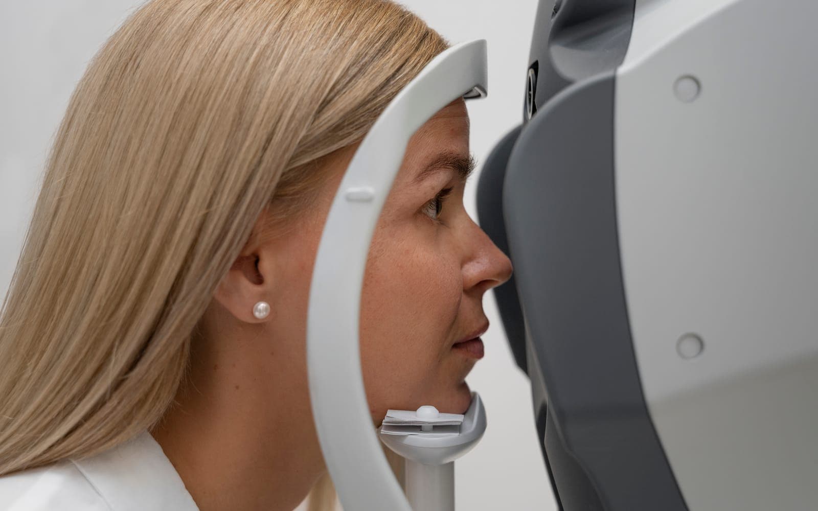
The basis of diagnosis is a retinal examination. Dilating the pupil and examining the retina in detail is important for determining the presence and severity of retinopathy.
In addition, different imaging methods may be used depending on the patient's retinal findings.
OCT (Optical Coherence Tomography) images the layers of the retina in detail and helps assess fluid accumulation and edema, particularly in the macular area.
Fundus photography allows changes in the retinal vessels, hemorrhages and other lesions to be documented and compared over time.
FFA (Fundus Fluorescein Angiography) shows the passage of dye through the vessels and can help assess leakage, vessel blockage and some areas of neovascularization.
Methods such as wide-field retinal imaging may also help evaluate peripheral retinal findings in suitable patients.
Not all of these tests are needed for every patient. Which imaging method is required is determined by the findings of the retinal examination.
How is diabetic retinopathy treated?
There is no single treatment for diabetic retinopathy that applies to everyone. The treatment decision is made by evaluating the active retinal pathology in each eye separately. This approach is also specifically emphasized in the current NICE guideline.
In milder cases of retinopathy, regular retinal monitoring and systemic control of diabetes come first. Managing blood sugar, blood pressure and blood lipids appropriately helps reduce the risk of retinopathy developing or progressing.
When findings such as retinal edema, advanced vascular changes or new vessel formation appear, different treatment options may come into consideration:
- Anti-VEGF injections: They may be used particularly for diabetic macular edema that affects vision and in some cases of proliferative retinopathy.
- PRP (Panretinal Photocoagulation)*: It is applied particularly in proliferative diabetic retinopathy to suppress the formation of abnormal new vessels and reduce the risk of serious vision loss.
- Macular laser: It may be used to reduce leakage in the macula, particularly in certain cases of diabetic macular edema.
- Intravitreal steroid treatments: In some cases of diabetic macular edema, they may be considered as an alternative to, or in addition to, other treatments.
- Vitrectomy*: Surgical treatment may be required for advanced vitreoretinal complications such as vitreous hemorrhage or tractional retinal detachment.
These treatments are not simple alternatives to one another. For example, PDR and DME are not the same thing and both can be present in the same patient. While approaches such as PRP or anti-VEGF are considered in PDR, the choice of treatment in DME may vary depending on factors such as whether the edema involves the center of the macula, how much vision is affected and other retinal findings.
Therefore, generalizations such as "if there is diabetic retinopathy, laser is done" or "if there is retinopathy, an injection is needed" are not correct. Treatment is determined according to the active problem in the retina.
When should an eye exam be done if you have diabetes?
The timing of eye exams for diabetic retinopathy depends on the type of diabetes, the duration of the disease and the current retinal findings.
According to ADA 2026, in adults the first comprehensive eye examination should generally be performed 5 years after the onset of type 1 diabetes and at the time of diagnosis in type 2 diabetes.
In some people who have no retinopathy and have adequate glycemic control, the screening interval may be extended to 1–2 years after one or more normal annual examinations. In contrast, if retinopathy of any degree is present, retinal evaluation at least once a year is needed, and more frequent follow-up is needed if the disease is progressing or threatens vision.
These intervals do not represent a fixed schedule for everyone. In particular, existing retinopathy, problems with diabetes control, pregnancy or vision-threatening retinal findings may require the follow-up frequency to be adjusted.
Can vision loss be prevented in diabetic retinopathy?
Early detection of diabetic retinopathy, timely treatment and good systemic management of diabetes play an important role in reducing vision-threatening complications.
However, the goal of treatment is not always to fully restore vision that has already been lost. Depending on the treatment, the goal may be to preserve current vision, to improve visual function by reducing edema in the macula, or to prevent serious complications such as hemorrhage and retinal detachment from developing.
Therefore, in diabetic retinopathy the most valuable approach is to detect retinal changes before they threaten vision, rather than seeking treatment after vision loss has occurred.
It should not be forgotten that diabetes control is also part of this process. Reaching blood sugar targets, and also managing blood pressure and lipid levels appropriately, helps reduce the risk of retinopathy developing or progressing.
Frequently Asked Questions
Can diabetic retinopathy be present in one eye but not the other?
Yes. The severity of retinal findings does not have to be the same in both eyes. For this reason, the two eyes are evaluated separately, and their follow-up or treatment needs may differ.
Are diabetic retinopathy and diabetic macular edema the same thing?
No. Diabetic retinopathy is the general term for diabetes-related changes in the retinal vessels. Diabetic macular edema is a condition in which fluid accumulates in the macular area as a result of increased vascular permeability. DME can develop at different stages of retinopathy.
What is the difference between diabetic retinopathy and cataract?
They are different diseases. A cataract is the loss of transparency of the eye's natural lens. Diabetic retinopathy, on the other hand, affects the retina and the retinal vessels. Although diabetes can affect the risk of developing both conditions, their location and treatments are different.
Does lowering blood sugar quickly reverse retinopathy?
Good glycemic control is important for reducing the risk of retinopathy developing and progressing; however, it does not mean that existing retinal damage will automatically disappear. Particularly in diabetes that has been poorly controlled for a long time, treatment goals and glycemic changes should be monitored by a physician.
Can diabetic retinopathy be completely cured?
It is not always possible for existing damage in the retina to be fully reversed. However, with appropriate follow-up and treatment, the progression of the disease can be slowed, the risk of vision loss can be reduced, and in some cases an improvement in visual function can be achieved.
Does pregnancy affect diabetic retinopathy?
Yes. Particularly in people with diabetes who already have retinopathy before pregnancy, it is important to plan retinal monitoring during pregnancy. The course of retinopathy and the need for treatment during pregnancy are evaluated separately, and the balance of benefit and risk for the mother and the fetus is taken into account when choosing treatment.
Is an eye examination enough for diabetic retinopathy?
A retinal examination is the basis of diagnosis; however, when necessary, the evaluation can be expanded with OCT, fundus photography, FFA or other retinal imaging methods. Which tests are needed is determined by the patient's retinal findings.
Is laser always done if there is diabetic retinopathy?
No. Laser treatment is not used at every stage of retinopathy or in every patient. PRP in particular is considered in proliferative diabetic retinopathy when certain retinal findings are present. In diabetic macular edema, the choice of treatment varies according to the characteristics of the edema and the extent to which vision is affected.
-ADA 2026 – Retinopathy, Neuropathy, and Foot Care
-American Academy of Ophthalmology – What is diabetic retinopathy?
Scientific references
- Wang W, Lo ACY. Diabetic Retinopathy: Pathophysiology and Treatments. Int J Mol Sci. 2018. PMID: 29925789
- Stitt AW, Curtis TM, Chen M, et al.. The progress in understanding and treatment of diabetic retinopathy. Prog Retin Eye Res. 2016. PMID: 26297071
- Tan TE, Wong TY. Diabetic retinopathy: Looking forward to 2030. Front Endocrinol (Lausanne). 2022. PMID: 36699020