Short answer
Topography maps the surface shape of the cornea, and pachymetry measures its thickness. Because laser surgery removes or reshapes corneal tissue, these tests are done beforehand to check that the cornea is thick and regular enough, and to look for problems such as hidden keratoconus that could make surgery unsafe.
Topography and pachymetry are done before laser eye surgery because the laser works by changing a very thin amount of corneal tissue, and the cornea must be able to tolerate that. Topography maps the curvature of the cornea, while pachymetry measures its thickness. Together they provide the basic information needed to plan surgery safely.
What are topography and pachymetry?
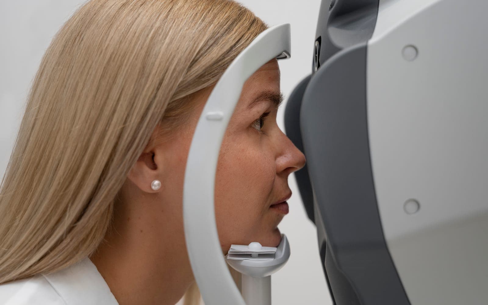
Topography is a non-contact scan that does not hurt that shows the curvature of the front surface of the cornea as a coloured map. Pachymetry is the measurement of corneal thickness. Most of today's devices provide both at the same time.
Older topography looked at the front surface alone, whereas newer systems show more. Scheimpflug imaging measures both the front and back surfaces of the cornea and builds a three-dimensional model. Devices such as the Pentacam and Galilei are used for this purpose, so a thickness map can be read together with elevation data.
Why are these measurements needed before laser surgery?
Laser corrects the prescription by changing the shape of the cornea, which involves removing tissue or reshaping it within the cornea. If the cornea is thin, weak or irregular, it may lose structural strength after surgery. The purpose of the measurements is to spot this risk before the operation.
These tests look for answers to questions such as:
- Is the cornea thick enough for the planned correction?
- Is the surface regular, or is there a bulge or thinning that suggests keratoconus?
- Is there a change on the back surface that does not show on the front-surface map?
- Do the prescription and astigmatism match the shape of the cornea?
How are ectasia risk and hidden keratoconus screened?
Ectasia is a weakening of the cornea with forward bulging, and it is one of the most feared complications of refractive surgery. Its causes include genetic susceptibility and external factors such as eye rubbing. Removing a large amount of tissue, as in very high prescriptions, may also add to the risk.
In one reported series of roughly thirty thousand eyes, post-LASIK ectasia occurred in 0.033%. The same review notes that this is lower than the earlier reported figure of 0.66%, and that better screening contributed to the fall. It is still accepted that, in rare cases, ectasia can appear despite thorough screening.
Early, mild keratoconus is difficult to catch. An eye that looks normal on the front-surface map can still look suspicious on back-surface data. You can read more in hidden keratoconus and laser surgery.
What do epithelial maps and biomechanical tests add?
The thickness map of the epithelium, the outermost layer of the cornea, can be produced with optical coherence tomography. Reviews state that it helps identify candidates at higher risk of complications and assists surgical planning.
Biomechanical devices such as the Corvis ST and the Ocular Response Analyzer look not at the shape but at the mechanical strength of the cornea. Imaging systems can reveal structural changes but cannot measure stability, and biomechanical testing tries to fill that gap. Even so, reviews stress that no universally accepted screening protocol or cut-off value exists in this area. For that reason, results are judged by the surgeon as a whole rather than by a single number.
How can you help the results be reliable?
Contact lenses can temporarily alter the shape of the cornea and distort the map, so you will be asked to leave them out for a period before measurement. The length of time is explained in stopping contact lenses before laser surgery. Avoiding eye rubbing is also important for both measurement accuracy and corneal health.
Measurements can be repeated when needed. If results are inconsistent or borderline, the doctor may add another imaging method or ask for a follow-up check after some time. This is a normal part of the process.
What do the results mean?
A borderline corneal thickness does not automatically rule out laser; the discussion turns to which method needs less tissue. This is addressed in is laser surgery really risky with a thin cornea. If results are not suitable for laser, options such as an intraocular lens may be considered. For the wider picture, see what are LASIK and FemtoLASIK.
If you have sudden vision loss, severe pain or a curtain-like shadow in your vision before or after surgery, see an eye doctor without delay.
Frequently Asked Questions
Does topography hurt?
No. The device does not touch your eye; you are simply asked to look at a point. It usually takes a few minutes and may not need drops.
What if my corneal thickness is not enough?
This does not always mean laser is impossible. The doctor considers a method that needs less tissue, or a different option, depending on your prescription and corneal structure.
Are these tests needed for every type of laser?
Yes. Corneal assessment is carried out before all procedures that work on the cornea, including LASIK, PRK and SMILE. The details of the measurements may differ between methods.
If the measurements are normal, is the risk of ectasia zero?
No. Screening lowers the risk considerably but does not remove it, which is why follow-up checks after surgery matter as well.
Scientific references
- Motlagh MN, Moshirfar M, Murri MS, et al.. Pentacam Corneal Tomography for Screening of Refractive Surgery Candidates: A Review of the Literature, Part I. Med Hypothesis Discov Innov Ophthalmol. 2019. PMID: 31598520
- Moshirfar M, Motlagh MN, Murri MS, et al.. Advances in Biomechanical Parameters for Screening of Refractive Surgery Candidates: A Review of the Literature, Part III. Med Hypothesis Discov Innov Ophthalmol. 2019. PMID: 31598522
- Salomão MQ, Hofling-Lima AL, Lopes BT, et al.. Role of the corneal epithelium measurements in keratorefractive surgery. Curr Opin Ophthalmol. 2017. PMID: 28399067
- Ambrósio R. Post-LASIK Ectasia: Twenty Years of a Conundrum. Semin Ophthalmol. 2019. PMID: 30664391