Short answer
Hidden keratoconus is a condition that causes no symptoms yet but may show early signs on corneal measurements. Because it is linked to corneal ectasia, a bulging of the cornea after laser surgery, it is generally treated as an important warning sign before laser. The decision rests on topography and tomography findings and is made by your doctor.
Hidden keratoconus is a condition in which the cornea shows early signs on measurement but causes no symptoms yet, and it is considered an important warning sign for laser eye surgery. Laser is generally not recommended in eyes with this condition because it may raise the risk of corneal bulging after surgery. Not every suspicious measurement means keratoconus, however; the assessment relies on examination and detailed corneal imaging.
What is hidden keratoconus?
Keratoconus is a disease in which the cornea progressively thins and bulges forward. It leads to irregular astigmatism and reduced vision. According to reviews, it usually begins in the second and third decades of life and can progress until the fourth decade. The disease can affect both eyes, but reviews describe it as bilateral and asymmetric, meaning the two eyes may not be affected at the same speed or to the same degree. For this reason, even if one eye looks normal, the measurements of the other eye are assessed separately. In the early stages, known as subclinical or forme fruste keratoconus, there may be no symptoms and glasses may still give good vision. For this reason a person may not know they have it. Diagnosing the early stages can be challenging even for specialists.
Why does it matter for laser surgery?
Laser corrects the prescription by removing corneal tissue. If the cornea is already biomechanically weak, the weakness can progress after surgery and lead to ectasia, a bulging of the cornea. Post-LASIK ectasia is described as one of the serious complications of refractive surgery, appearing as corneal thinning and bulging along with increasing myopia and astigmatism. According to reviews, ectasia can be associated with factors that heavily affect the structure of the cornea, such as treatment of high myopia, or with reduced biomechanical strength before surgery. Genetic factors and environmental factors such as eye rubbing are also listed among the risks. This is why screening before surgery is so important.
How is hidden keratoconus detected?
A single parameter may not be enough for diagnosis. The following tests are usually assessed together:
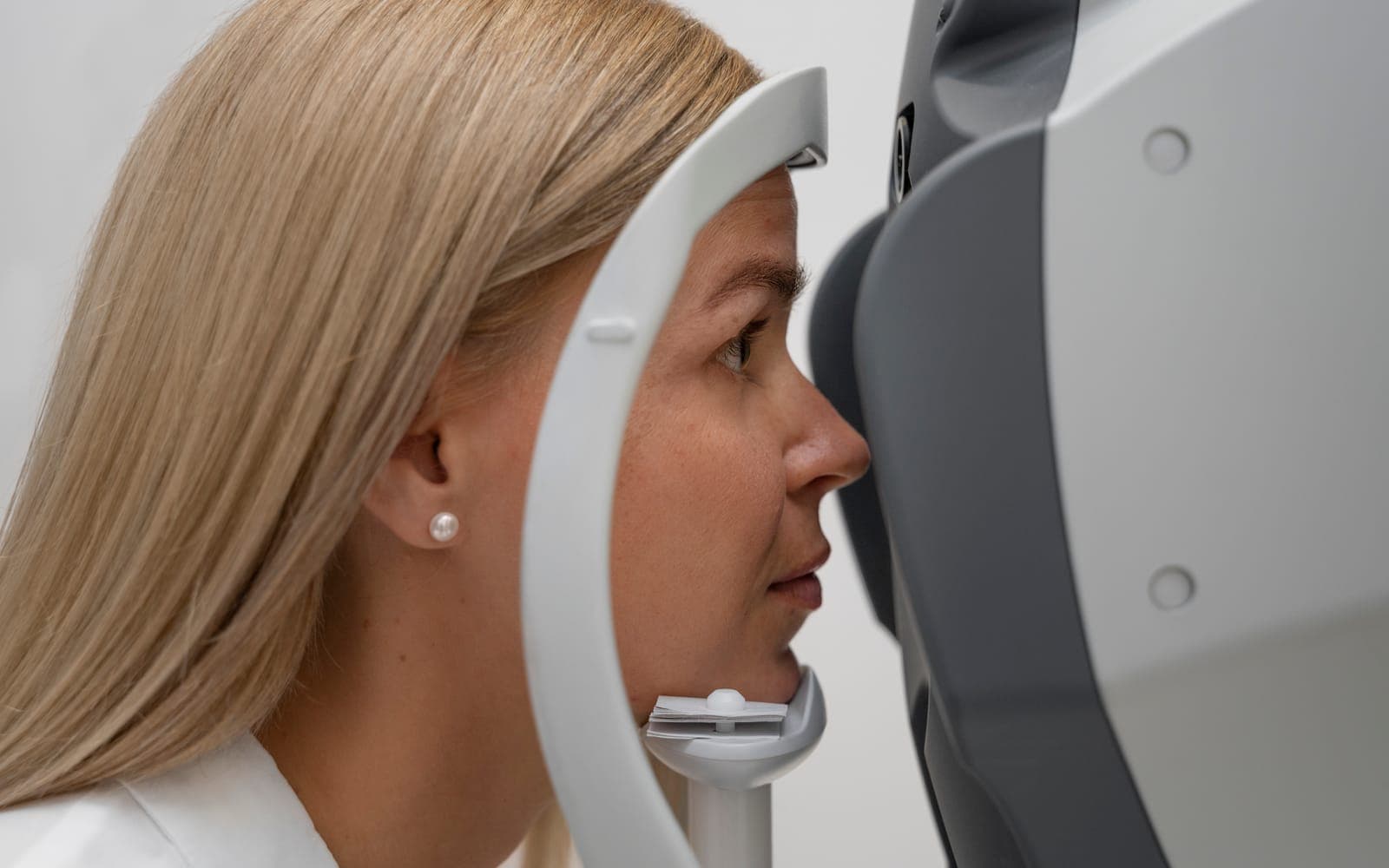
- Corneal topography and tomography, which show the curvature of the front and back surfaces.
- A corneal thickness map (pachymetry) and the distribution of thickness.
- Devices that measure corneal biomechanics and methods based on optical coherence tomography.
- Questions about family history, eye rubbing and risk factors such as allergy and eczema.
Reviews report that tomography, which provides data on the back surface and thickness distribution, is used to detect early or subclinical disease and to screen laser candidates. Artificial intelligence-assisted methods are also being studied, but they do not replace assessment by an experienced doctor. For details on these tests, see our article on topography and pachymetry before laser surgery.
What happens if hidden keratoconus is found?
Early detection matters because early intervention can halt progression and reduce the risk of ectasia and the need for a corneal transplant. When hidden keratoconus is suspected, the following approaches are commonly considered:
- Laser surgery is avoided, or a more careful follow-up plan is made.
- Measurements are repeated at intervals to see whether the disease is progressing.
- If it progresses, corneal cross-linking (CXL) may be used. CXL uses riboflavin and ultraviolet A light to stiffen the cornea.
- Stopping eye rubbing is advised.
Your doctor decides which option suits you based on your findings.
Are there alternatives if I cannot have laser?
Yes. Glasses, contact lenses or intraocular lenses may be considered. For higher prescriptions see our article on options for high myopia, and for phakic lenses see the phakic intraocular lens (ICL). The appropriate method becomes clear after examination.
How is ectasia treated if it develops?
Corneal cross-linking (CXL) is a method used to strengthen the cornea in progressive keratoconus and ectasia. In the original "Dresden" protocol, introduced about twenty years ago, the cornea is saturated with riboflavin and then exposed to ultraviolet A light at 3 mW/cm2 for 30 minutes. This creates cross-links between the fibres of the cornea and stiffens the tissue. The original protocol requires a minimum corneal thickness of 400 microns to protect the endothelium. Accelerated protocols that shorten the procedure, and approaches that do not remove the surface layer, have since been developed. Your doctor decides which one suits you. CXL aims to halt progression; it does not always restore lost vision, and like any procedure it has risks.
How can I protect myself?
Family history, eye rubbing, allergy and eczema are listed among the risk factors for keratoconus. For this reason, try not to rub your eyes, have allergy symptoms treated, and have an examination if your glasses prescription changes quickly. If you are considering laser, tell your doctor openly about your family history and habits.
Frequently Asked Questions
Does a borderline topography mean I have keratoconus?
No. A borderline value does not make a diagnosis on its own. Tomography, thickness and clinical findings are assessed together, and follow-up measurements are sometimes needed.
Does hidden keratoconus cause symptoms?
It usually causes none, or only mild changes in prescription. That is why screening before laser is important.
Does eye rubbing cause keratoconus?
Eye rubbing is one of the reported risk factors for keratoconus and ectasia. If you rub your eyes often, tell your doctor.
What should I do if my vision suddenly worsens?
If you have sudden vision loss, severe eye pain or a sense of a curtain over your vision, see an eye doctor without delay.
Scientific references
- Al Bdour M, Sabbagh HM, Jammal HM. Multi-modal imaging for the detection of early keratoconus: a narrative review. Eye Vis (Lond). 2024. PMID: 38730479
- Ambrósio R. Post-LASIK Ectasia: Twenty Years of a Conundrum. Semin Ophthalmol. 2019. PMID: 30664391
- Ghemame M, Charpentier P, Mouriaux F. Corneal topography in clinical practice. J Fr Ophtalmol. 2019. PMID: 31727328
- Hafezi F, Kling S, Hafezi NL, et al.. Corneal cross-linking. Prog Retin Eye Res. 2025. PMID: 39681212