Short answer
OCT-A is a non-invasive imaging method that visualizes the vascular network of the retina and choroid and blood-flow-related changes without injecting contrast dye. It helps assess vascular structure especially in diabetic retinopathy, retinal vascular occlusions and age-related macular degeneration. When vessel leakage needs to be examined, fluorescein angiography (FFA) may still be required.
OCT-A (Optical Coherence Tomography Angiography) is a non-invasive eye imaging method used to visualize the vascular network in the retina and choroid and changes related to blood flow. It helps evaluate the vascular structure especially in diabetic retinopathy, retinal vascular occlusions and age-related macular degeneration.
While OCT* evaluates the structural features of the retina and macular tissues in detail, OCT-A focuses on the microvascular structure in these areas.
What Is OCT-A?
OCT-A is an advanced imaging method that allows the vascular network in the retina and choroid to be visualized. By analyzing signals related to blood flow without requiring a contrast agent to be given through a vein, it helps evaluate vascular structures in different layers of the retina and choroid.
Thanks to this feature, it can be used especially to examine the small vessels and microvascular changes in the back of the eye.
What Is the Difference Between OCT and OCT-A?
Although OCT and OCT-A use similar technological foundations, they focus on evaluating different information. OCT mainly shows the structural features of the retina, macula and other eye tissues, while OCT-A focuses on evaluating the vascular network and changes related to blood flow.
| Feature | OCT | OCT-A |
|---|---|---|
| What does it mainly show? | Structural features of eye tissues | The vascular network in the retina and choroid |
| Main purpose of use | Evaluating tissue layers and structural changes | Evaluating vascular structure and microvascular changes |
| Vascular assessment | Assessment through structural imaging | Focuses on imaging the vascular network |
| Contrast agent | Not required | Not required |
| Areas of use | Retina, macula, glaucoma and various eye diseases | Diabetic retinopathy, retinal vascular occlusions and macular-choroidal vascular changes |
| Does it completely replace FFA? | No | No; FFA may be needed especially when vascular leakage needs to be evaluated |
For this reason, OCT and OCT-A are not alternatives to each other but imaging methods that complement each other when needed.
How Does OCT-A Work?
OCT-A analyzes changes in the optical signals obtained from tissues and helps reveal vascular structures in which moving blood cells are present. In this way, the vascular networks at different depths of the retina and choroid can be evaluated separately.
No contrast agent injection is required during the examination. In the images obtained, vessel density, the arrangement of the vascular network and the microvascular changes seen in some diseases can be examined.
However, eye movements, imaging artifacts and image quality can affect the evaluation. For this reason, OCT-A findings are not evaluated alone but together with the eye exam and, when necessary, other imaging methods.
In Which Eye Diseases Is OCT-A Used?
OCT-A is used especially to evaluate vascular changes in the retina and macula. In which disease and for what purpose it will be used is determined according to the patient's clinical findings.
Diabetic Retinopathy*
In diabetic retinopathy, microvascular changes in the retinal vessels are among the important findings of the disease. OCT-A can help evaluate changes in vessel density and in the vascular network.
For this reason, it can be used together with other tests in the evaluation and follow-up of diabetes-related retinal changes.
Retinal Vascular Occlusions
In retinal vascular occlusions, microvascular changes can occur in areas where blood flow is impaired. OCT-A helps evaluate the vascular areas affected by the occlusion and the circulatory changes within the retina.
Age-Related Macular Degeneration
In neovascular age-related macular degeneration in particular, abnormal vessel formation can be seen in the macular region. OCT-A can help visualize these vascular networks and follow their changes over time.
Other Retinal and Choroidal Vascular Changes
OCT-A can also be used as a supportive imaging method in other retinal and choroidal diseases in which the vascular structure is clinically important. In some cases, detailed evaluation of changes in the vascular network may contribute to following the disease.
What Are the Advantages of OCT-A?
One of the important advantages of OCT-A is that vascular imaging can be performed without a contrast agent injection.
Its main advantages:
-
Helps evaluate the retinal and choroidal vascular network at different depths.
-
Allows microvascular changes to be examined.
-
Does not require a contrast agent injection.
-
Can be used in follow-up examinations when needed.
-
Can complement information obtained from other imaging methods such as OCT.
However, OCT-A has some limitations. Motion artifacts and low image quality during imaging can make evaluation difficult. In addition, OCT-A does not directly show vascular leakage.
Does OCT-A Replace Eye Angiography?
OCT-A does not replace FFA*', known as eye angiography, in every situation.
While OCT-A has an advantage in visualizing the vascular network and changes related to blood flow, it does not directly show vascular leakage. FFA, on the other hand, uses a fluorescein contrast agent given into a vein to help evaluate circulation and findings such as vessel permeability and leakage.
For this reason, while OCT-A may provide sufficient information in some patients, other imaging methods such as FFA may be needed, especially when vascular leakage needs to be evaluated.
When Is OCT-A Requested?
OCT-A may be requested when a detailed evaluation of the retinal or choroidal vascular structure is needed. It can be helpful especially in evaluating diabetic retinopathy, retinal vascular occlusions and vascular changes in the macular region.
However, whether OCT-A is needed is not determined by the name of the disease alone. The appropriate imaging method is chosen by evaluating the findings of the eye exam, the patient's complaints and other tests considered necessary.
Frequently Asked Questions
Is an OCT-A scan painful?
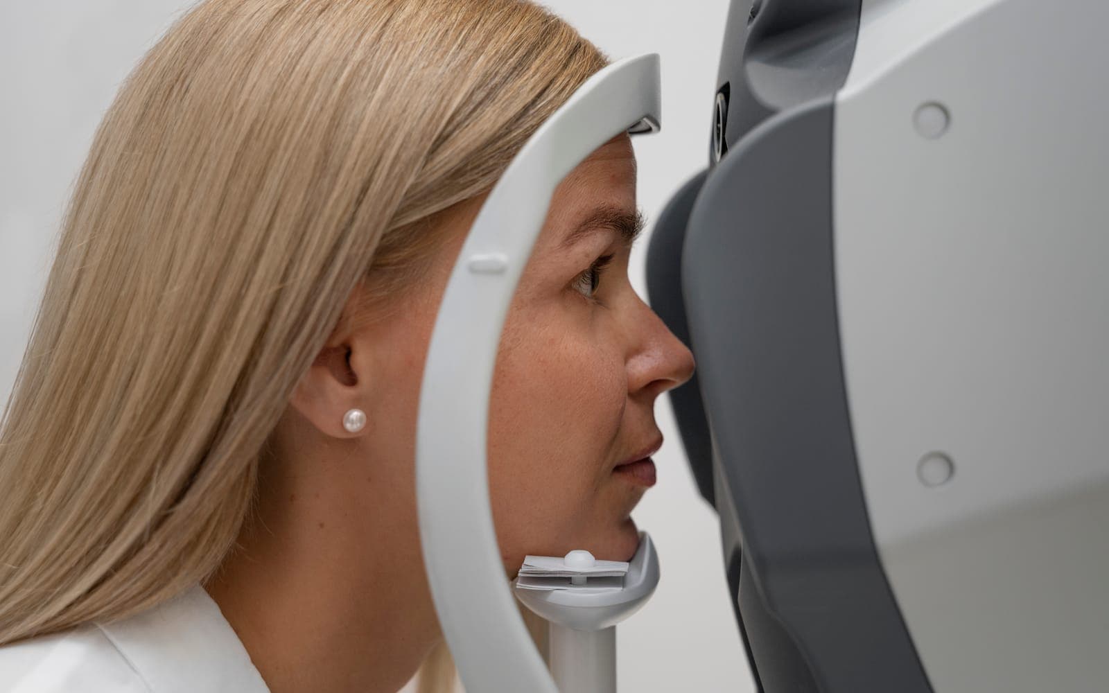
OCT-A is a non-invasive imaging method and does not require a contrast agent to be given through a vein. During imaging, it is enough for the patient to look at the designated point in front of the device.
Can a vascular occlusion be seen with OCT-A?
OCT-A can help evaluate microvascular changes caused by a retinal vascular occlusion and the affected vascular areas. Findings are interpreted together with other exam and imaging results.
Are OCT-A and OCT the same thing?
No. While OCT mainly evaluates the structural features of eye tissues, OCT-A focuses on the vascular network in the retina and choroid and changes related to blood flow.
Are OCT-A and eye angiography the same?
No. OCT-A images the vascular network without using a contrast agent. FFA, on the other hand, uses a fluorescein contrast agent given into a vein to help evaluate circulation and especially vascular leakage.
Does OCT-A diagnose on its own?
Not always. OCT-A can provide important imaging information, but results are evaluated together with the eye exam and, when necessary, other tests.
Scientific references
- Spaide RF, Fujimoto JG, Waheed NK, et al.. Optical coherence tomography angiography. Prog Retin Eye Res. 2018. PMID: 29229445
- Waheed NK, Rosen RB, Jia Y, et al.. Optical coherence tomography angiography in diabetic retinopathy. Prog Retin Eye Res. 2023. PMID: 37499857
- Sheth JU, Stewart MW, Narayanan R, et al.. Macular neovascularization. Surv Ophthalmol. 2025. PMID: 39222802